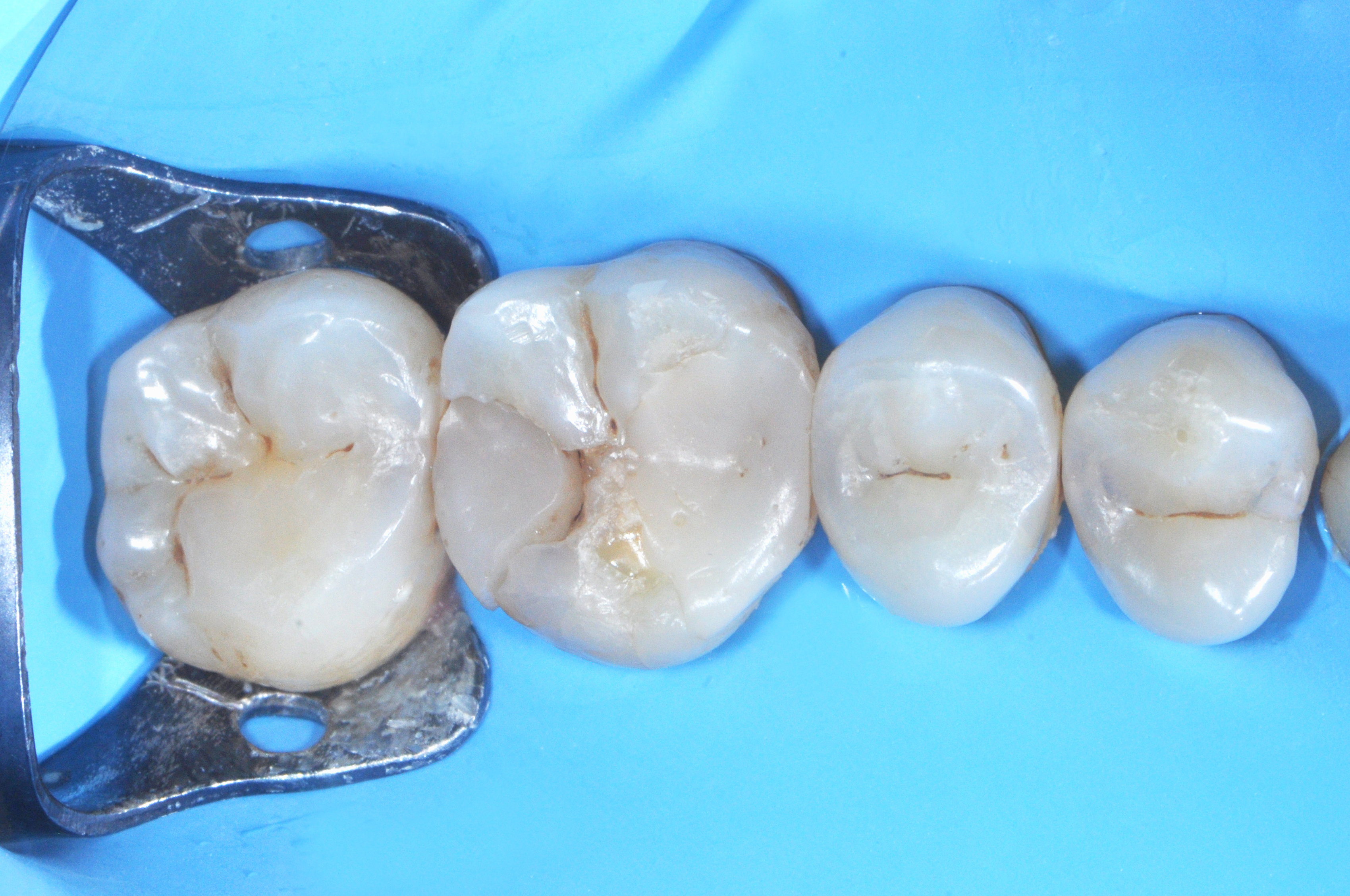
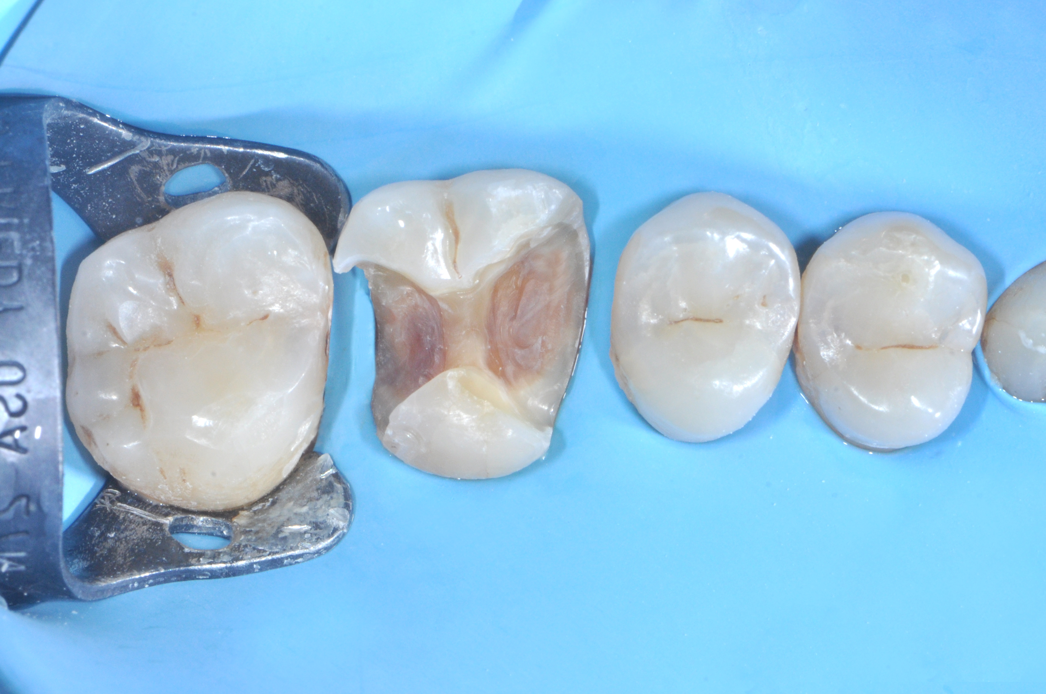
The patient presented for examination with a large, incongruous mesio-occluso-distal restoration on tooth 1.6, with clear evidence of secondary caries infiltration. Due to the patient’s limited financial resources, it was decided to manage the tooth with a direct freehand restoration (Figure 1).
The quadrant was isolated using a rubber dam (Figure 2).
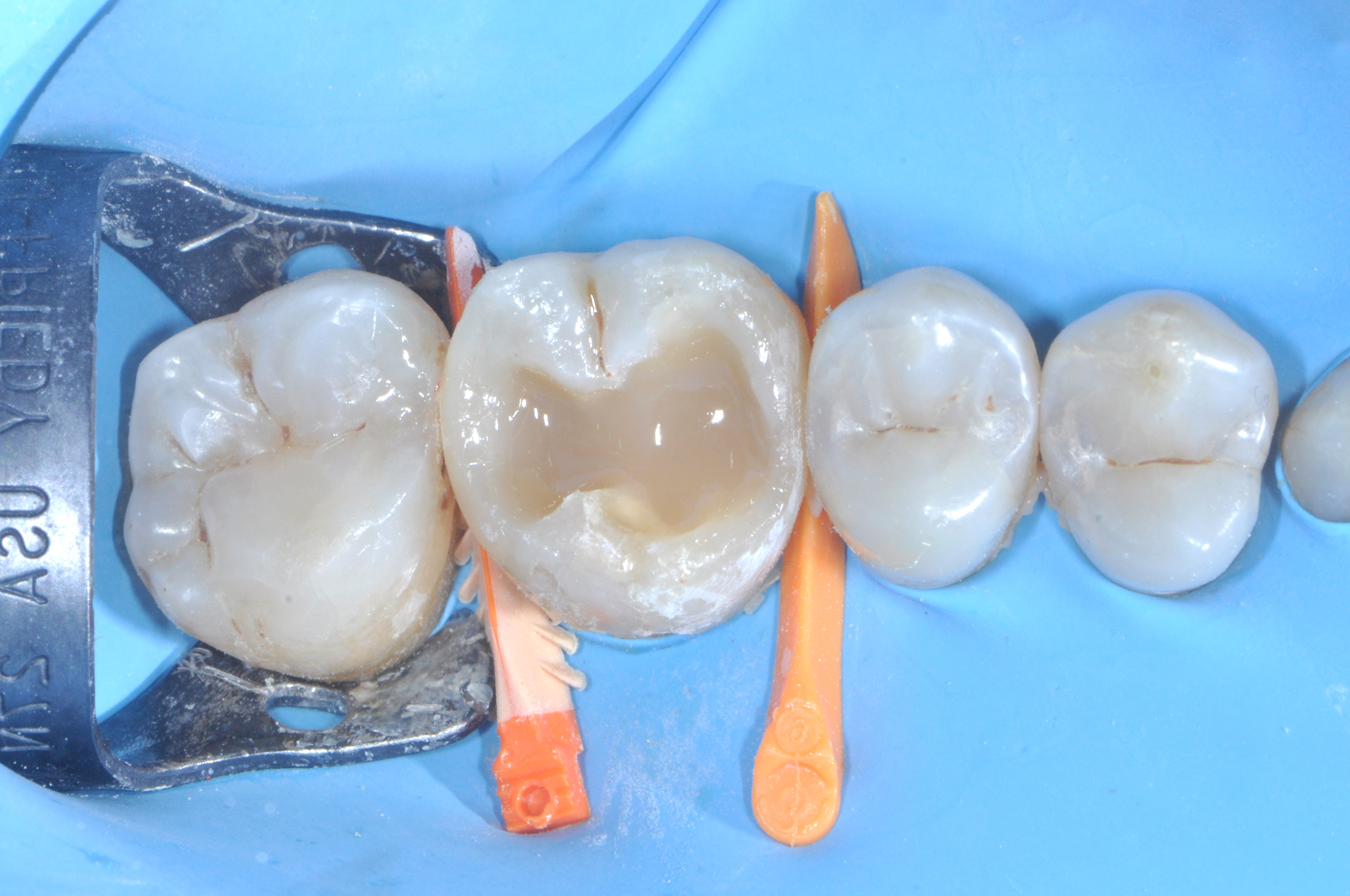
Removal of the old restoration and softened tooth structure revealed a very large MOD cavity with substantial loss of tooth substance. Not only were both proximal walls missing, but part of the palatal wall was also lost, particularly in the mesiopalatal area. The interproximal space between the cervical step of the boxes and the proximal walls of the adjacent teeth was notably wide (Figure 3)
|
|
|
| Figure 1. | Figure 2. | Figure 3. |
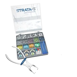
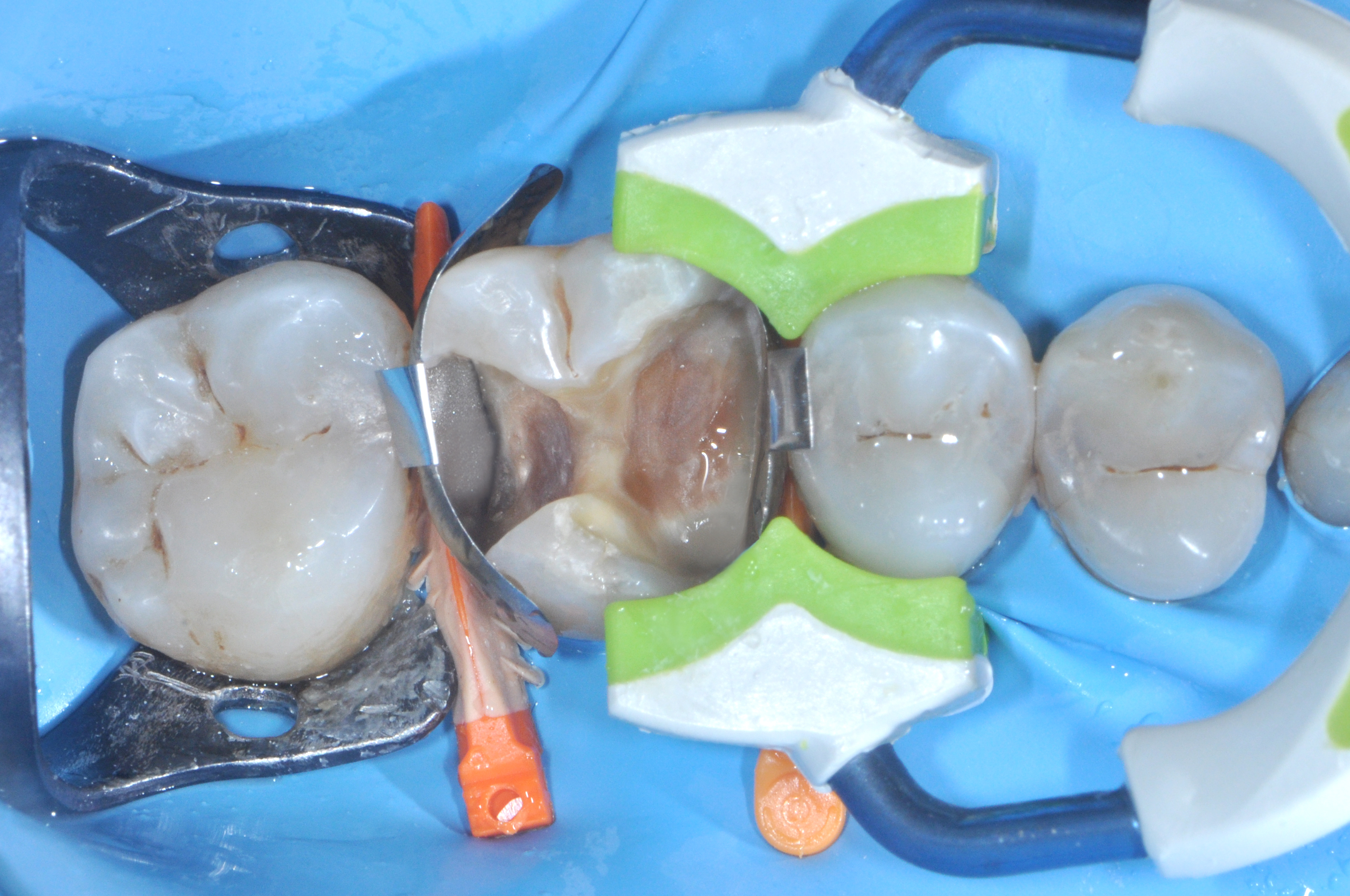
To obtain an optimal seal at the level of the two deep cervical margins, and given the significant distance between the cervical steps and the proximal walls of the adjacent teeth, it was decided to use a Firm Band matrix system. Thanks to its specific radius of curvature and pronounced convexity, this system allowed the recreation of proper emergence profiles without the need for additional instruments.
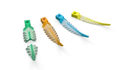
The concavity present at the distal cervical margin, which could have caused difficulties in adapting the matrix to the cervical profile, was managed using an orange 3D Fusion wedge. The expandable silicone fins allowed for a perfect seal of the gap between the matrix and the cervical margin in its central portion, corresponding to the anatomical concavity of that area. The loss of tooth structure affecting the palatal wall in its mesial portion was managed using a green Strata G separating ring. This ring was able to embrace the tooth perfectly, including the palatal aspect, ensuring optimal adaptation of the matrix to the axial walls of the mesial box (Figure 4).
|
| Figure 4. |
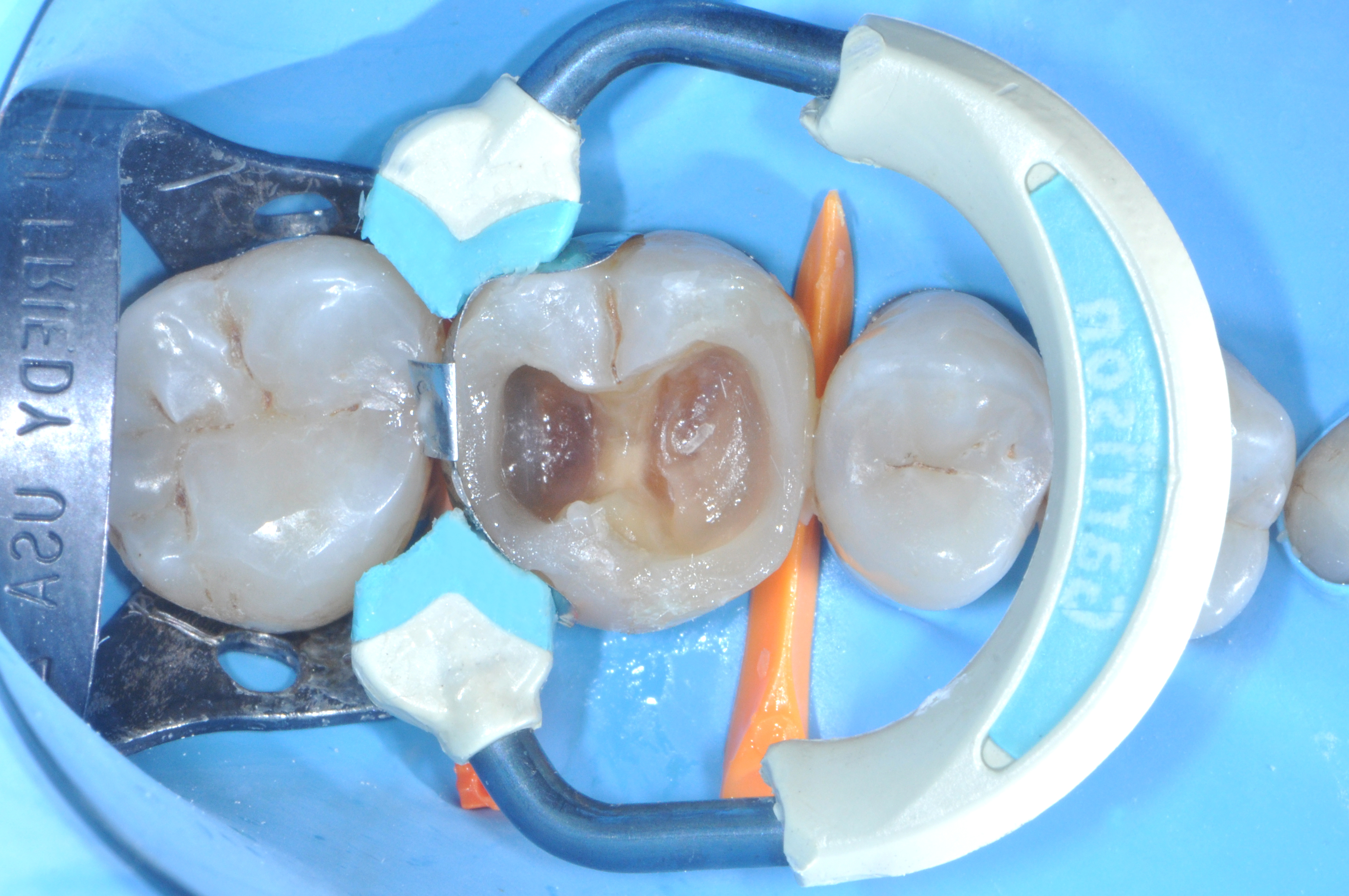
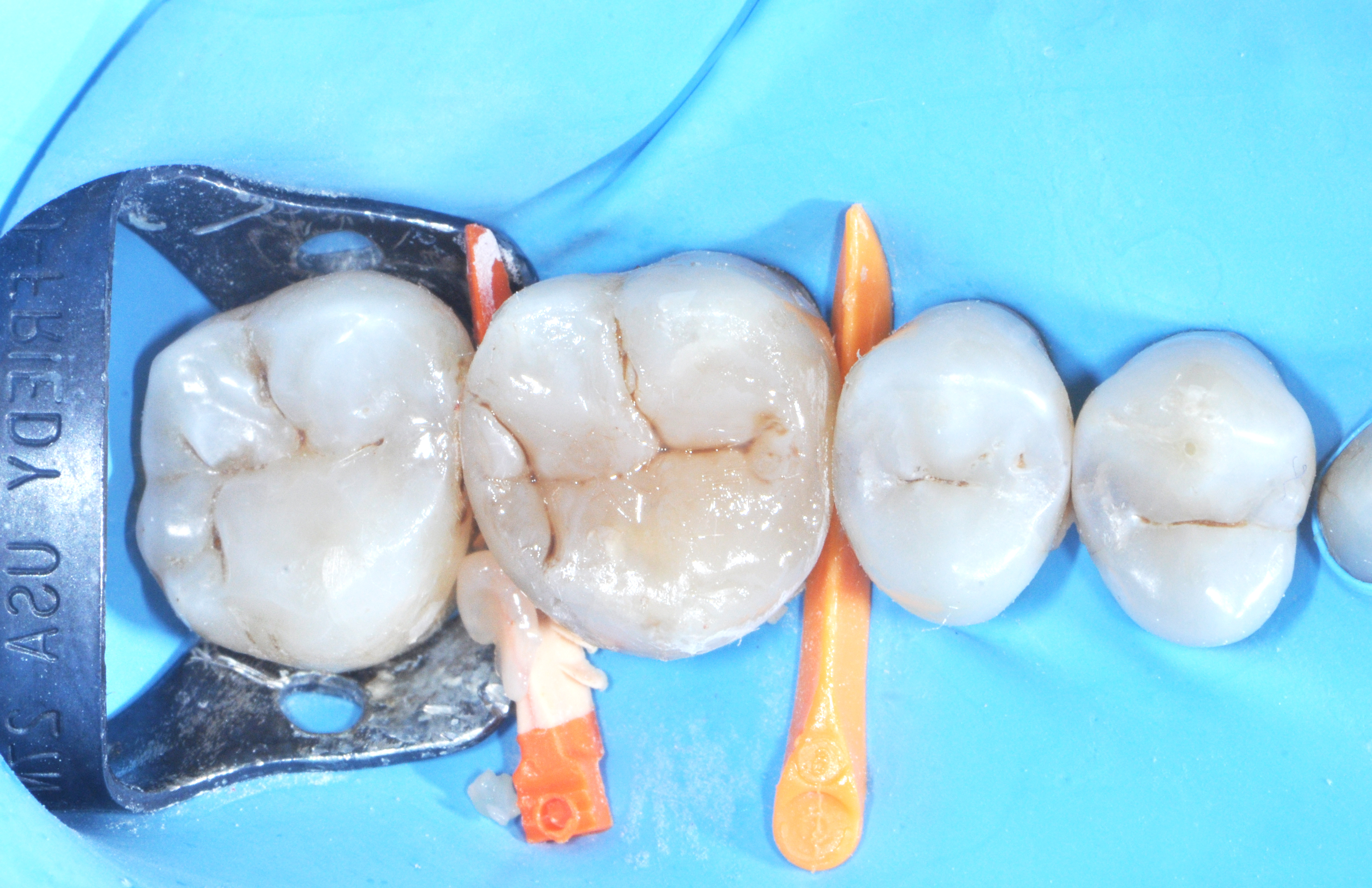
Once the mesial wall had been reconstructed, the mesial matrix was removed and a blue Strata G separating ring was adapted to manage the distal interproximal wall (Figure 5).
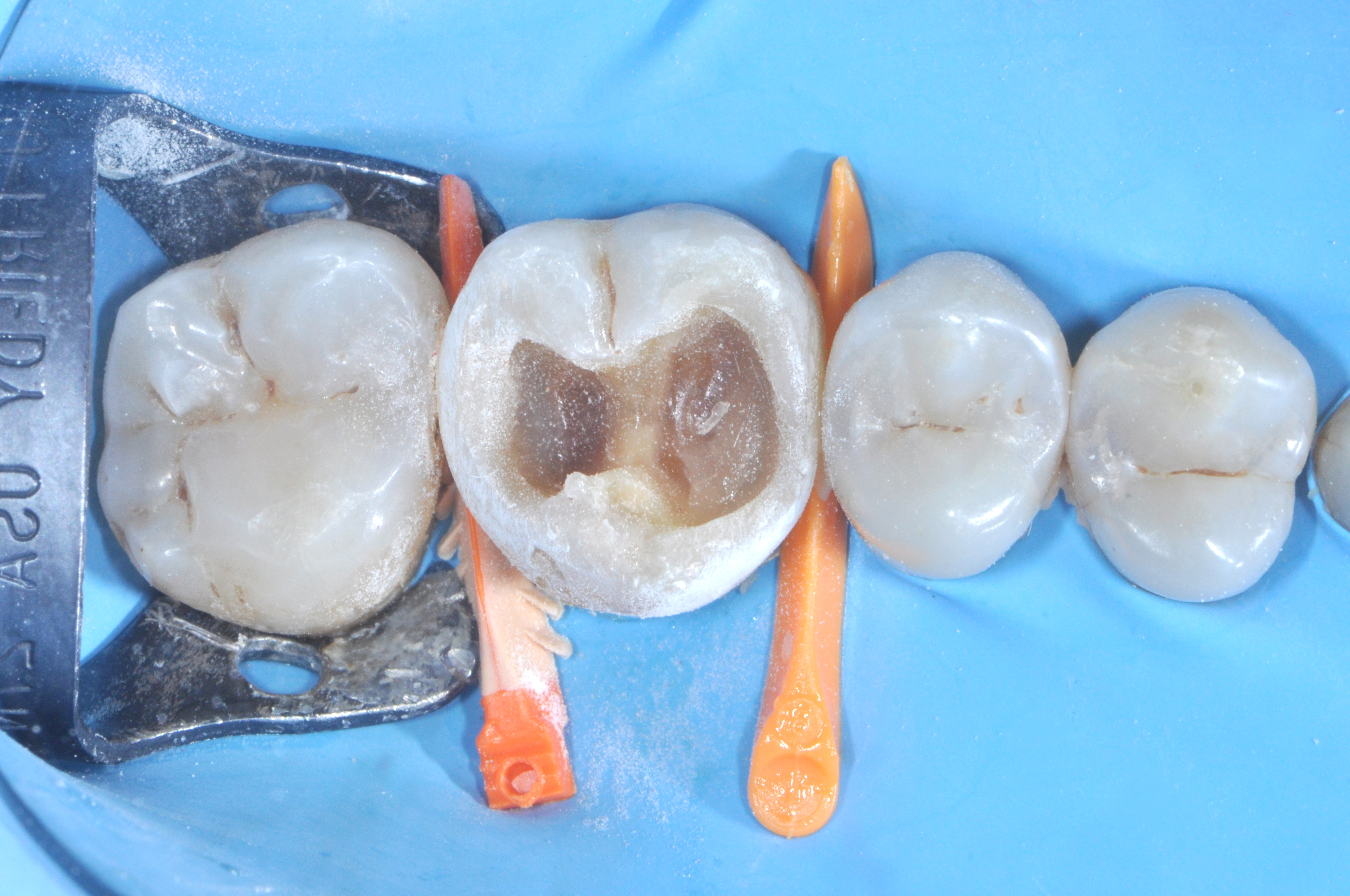
After reconstructing both interproximal walls, recontouring of the walls was performed using a coarse grit disc to reestablish a correct outline of the tooth prior to composite layering and occlusal anatomical modeling (Figure 6).
The remaining cavity was filled using horizontal layering with a highly filled flowable composite, leaving approximately 1.5mm of space at the occlusal level (Figure 7).
|
|
|
| Figure 5. | Figure 6. | Figure 7. |
Occlusal anatomical modeling was then performed cusp by cusp using a highly filled, nanofilled packable composite. The addition of brown and white stains allowed the restoration to integrate more biomimetically within the surrounding dentition, emulating the esthetics of the adjacent teeth (Figure 8).
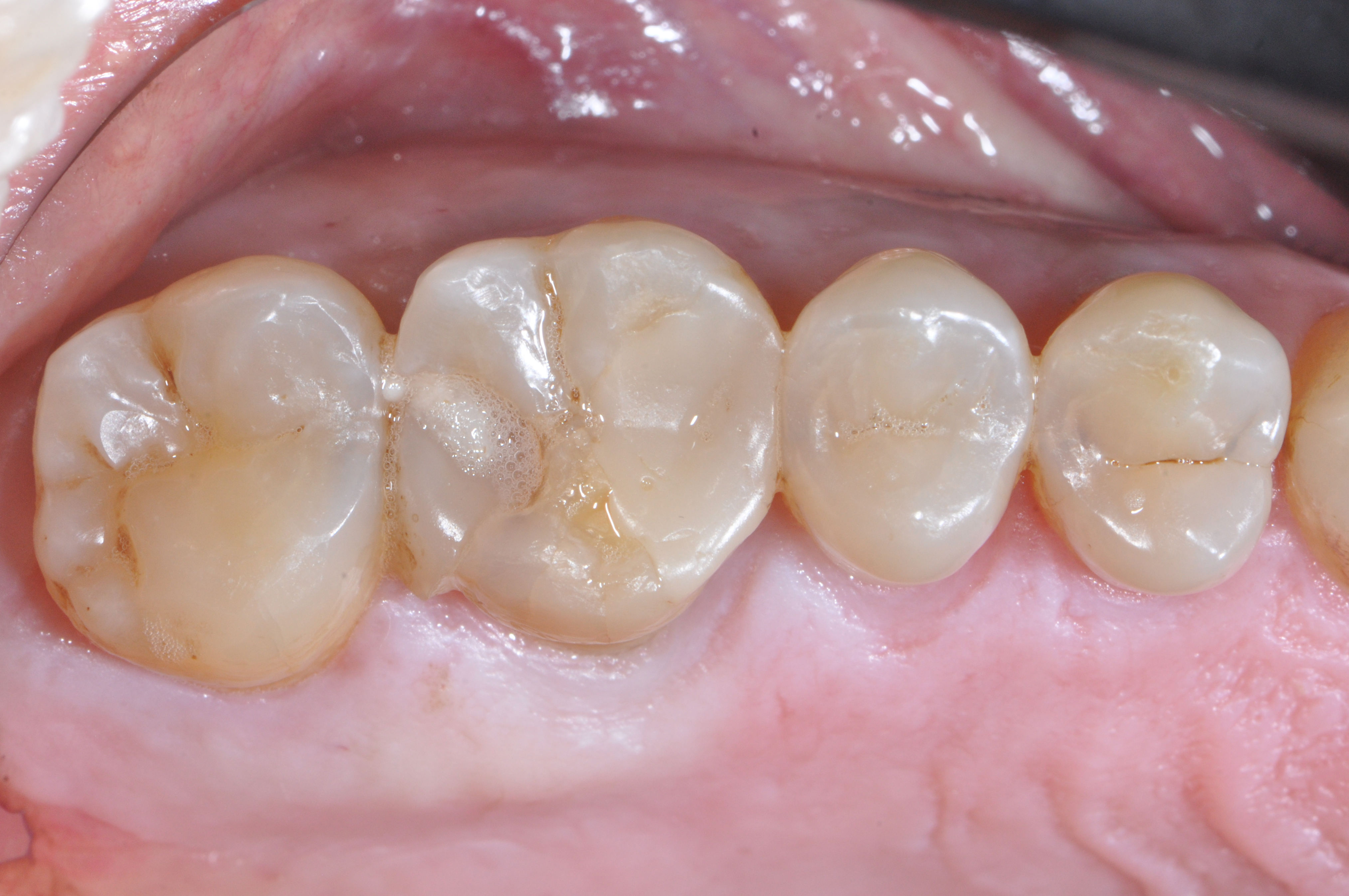
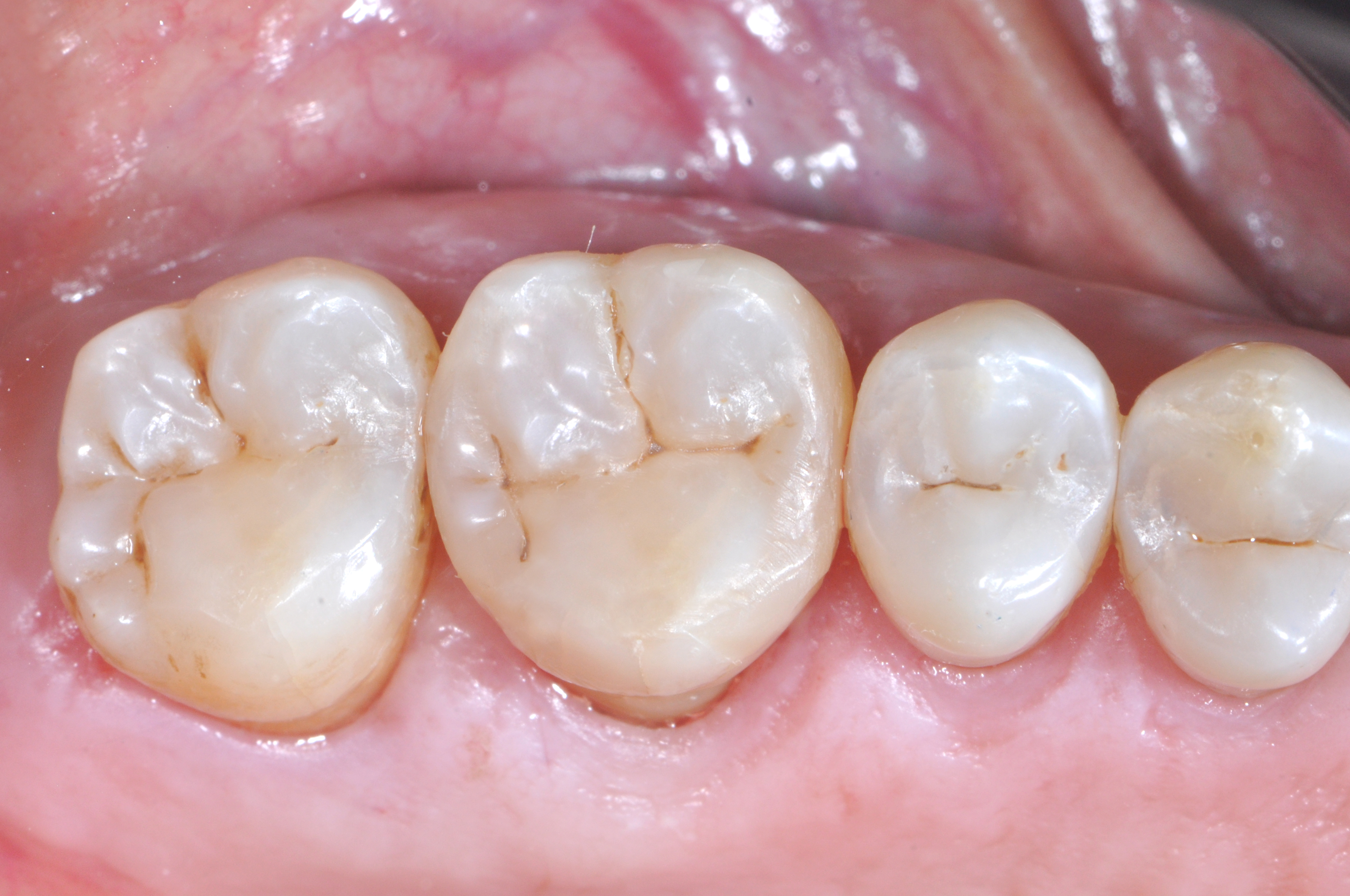
The restoration was finished and polished, with a 2 year follow up (Figure 9).
|
|
| Figure 8. | Figure 9. |