FREE SHIPPING on orders over $475
FREE SHIPPING on orders over $475 | No Code Needed
A fractured posterior cusp doesn’t always mean crown lengthening—or immediate endodontic treatment. When the margin is accessible, deep margin elevation can provide a conservative pathway from fracture to a predictable crown preparation.
A 69-year-old male presented to our practice as a new patient 12 months earlier. A comprehensive examination was completed, including a full-mouth radiographic series, intraoral photography, an iTero Wellness Scan and the Kois Diagnostic Opinion Form.
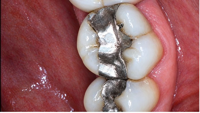
The patient was assessed as low risk for periodontal and dentofacial/esthetic disease, moderate risk for function and high risk biomechanically. A comprehensive treatment plan was presented. Except for periodontal therapy, the patient initially declined treatment of the biomechanically compromised teeth (Image 1).
|
|
|
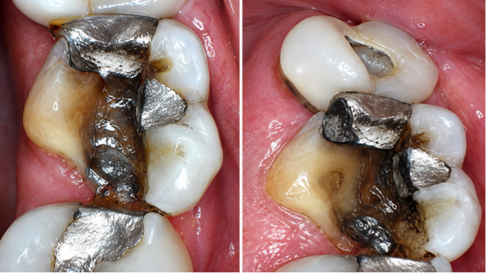
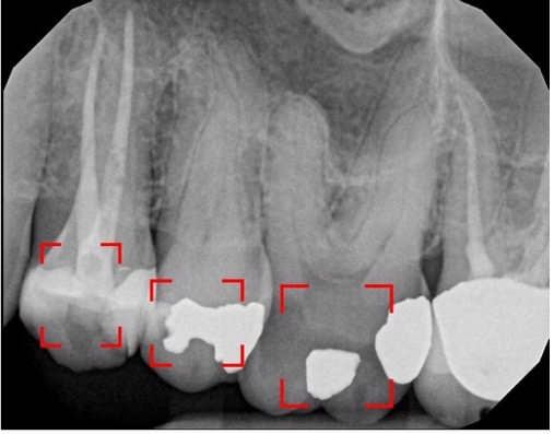
| Image 1: The initial presentation of tooth#26 | Image 2: One year later, he presented with an emergency: a fractured palatal cusp on tooth #26 |
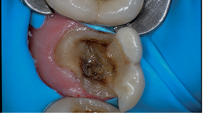
One year later, he presented with an emergency: a fractured palatal cusp on tooth #26 (Image 2).
Clinical and radiographic evaluation demonstrated a supra-osseous fracture, with no indication for endodontic treatment. Because the fracture remained accessible and did not require surgical exposure of the osseous crest, periodontal crown lengthening was not indicated at that time.
Teeth #24–27 had originally been treatment planned for indirect restorations. Following discussion of treatment options, the patient elected to proceed with deep margin elevation (DME) followed by crown treatment of teeth #24–27.
This case demonstrates the restorative sequence used to stabilize tooth #26 and establish a predictable, accessible margin prior to definitive crown preparation.
The Clinical Workflow
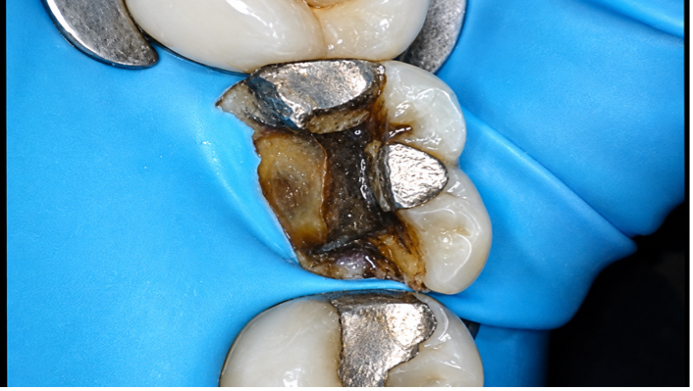
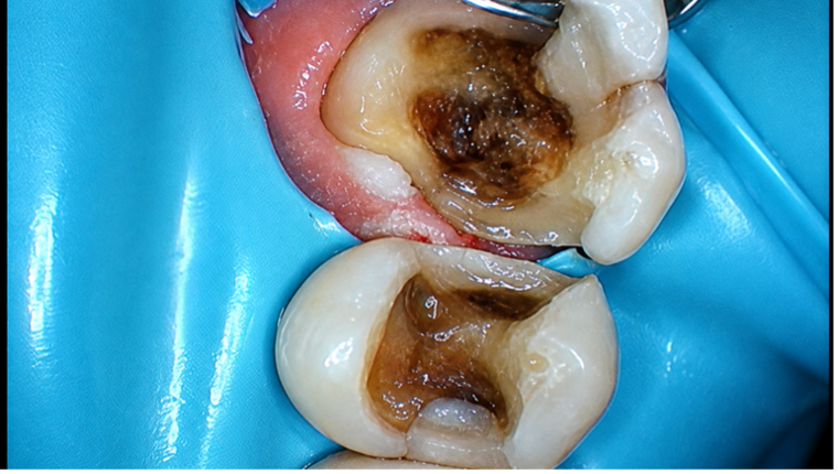
A rubber dam was placed before restorative treatment (Image 3).
|
|
| Image 3: A rubber dam was placed before restorative treatment |
For adhesive dentistry, isolation is not simply a matter of convenience. Contamination from saliva, blood or crevicular fluid can compromise the bonding procedure. In this case, rubber dam isolation also provided the visibility and access necessary to manage the deep cervical margin.
A 2021 Cochrane review found low-certainty evidence that rubber dam isolation may improve the short-term survival of some direct composite restorations, although the evidence at longer follow-up periods remains uncertain. ¹
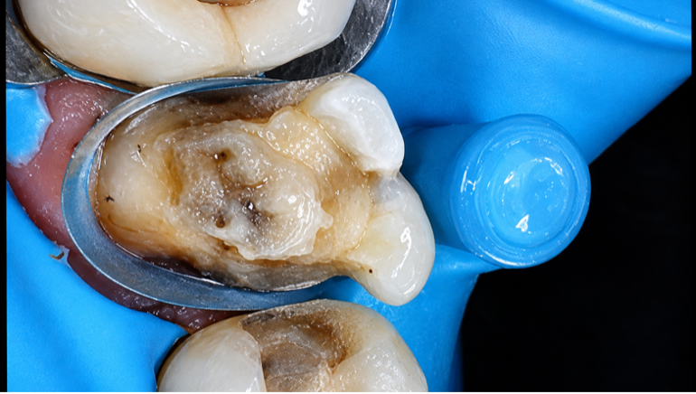
Existing amalgam was removed, and the final tooth preparation was completed. A soft-tissue laser (Blue Wave Diode Soft Tissue Laser, Clinician’s Choice) was then used to expose the clinical margin sufficiently to permit adaptation of the Garrison Dental Solutions DME Band (Images 4 and 5).
|
|
| Image 4: A soft-tissue laser was used to expose the clinical margin. | Image 5: Deep Margin Elevation band adapted to the cervical margin. |
The objective was not simply to "fill the box." The goal was to create a clean, accessible and controllable restorative margin.
The DME Band was carefully adapted to the cervical margin.
DME—also referred to as cervical margin relocation or proximal box elevation—uses bonded restorative material to relocate a deep cervical margin coronally. This can facilitate isolation, matrix adaptation, bonding and subsequent restorative procedures.²,³
Current reviews suggest that DME can be a conservative alternative to surgical crown lengthening in appropriately selected cases. However, the technique is highly dependent on three factors:
Importantly, much of the current DME evidence remains laboratory-based, with relatively limited long-term clinical data.²,³
Following preparation, the dentin was treated :
a. With pumice followed up with air abrasion (PrepStart Air Abrasion System, Danville Materials) to endure a clean surface ready for adhesive dentistry.
b. OptiBond FL (Kerr) a three-step total-etch/etch-and-rinse protocol.
After etching and rinsing, the dentin was re-wetted using G-5 (Clinician's Choice) before application of the adhesive.
The three-step etch-and-rinse technique remains one of the best-documented approaches to dentin adhesion. It separates the clinical functions of etching, priming and adhesive resin application, allowing each component to perform a specific role in creating the resin-dentin interface.⁴
OptiBond FL has demonstrated strong dentin-bonding performance in laboratory studies and long-term clinical evaluations of three-step adhesive systems have demonstrated favorable retention and marginal outcomes.⁵- ⁷
Evanesce BC (Clinician's Choice) was selected as the DME composite base.
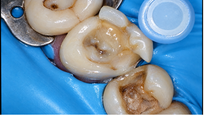
Its flowability allowed adaptation to the cervical margin and the internal aspect of the matrix. It was also selected due to its bio-adaptive technology and unlimited depth of cure. The material was carefully placed and polymerized to complete the margin elevation (Image 6).
|
|
| Image 6: Тhe material was placed and polymerized to complete the margin elevation. | Image 7: A more accessible coronal restorative margin achieved. |
The result was a new, more accessible coronal restorative margin. (Image 7)
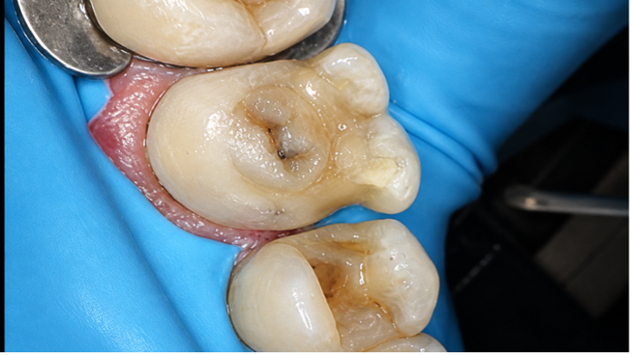
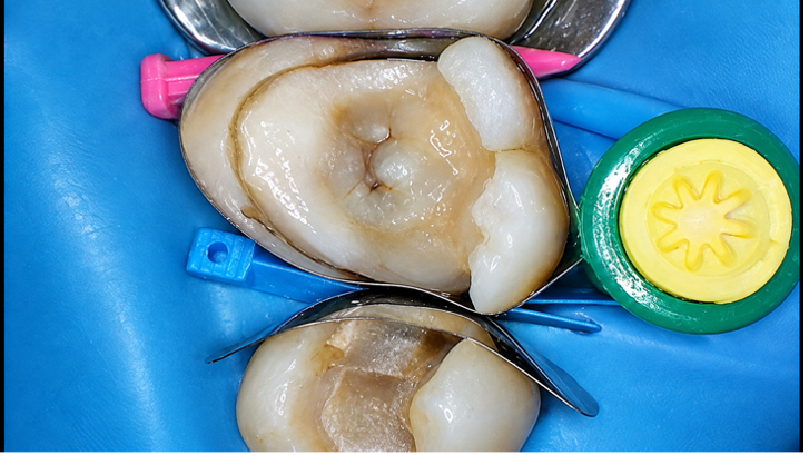
With the DME completed, tooth #26 and tooth #25 were restored using the Garrison Dental Solutions ReelMatrix and Quad Sectional Matrix system (Images 8 and 9).
Proper wedging was particularly important.
The matrix creates the contour; the wedge creates the seal.
A well-adapted wedge helps prevent composite from being extruded into the gingival sulcus and minimizes the need for difficult subgingival finishing. This becomes even more important when the cervical margin has been relocated with DME.
|
| Image 8: Tooth #26 and tooth #25 were restored using the Garrison Dental Solutions ReelMatrix and Quad Sectional Matrix system |
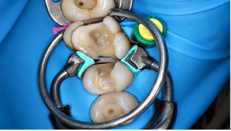
|
| Image 9: Tooth #26 and tooth #25 were restored using the Garrison Dental Solutions ReelMatrix and Quad Sectional Matrix system |
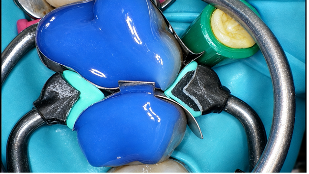
|
| Image 10: Tooth was treated with the total-etch technique using OptiBond FL, followed by G-5 rewetting agent |
7. Adhesive Protocol—Again
For the definitive restoration, the tooth was again treated with the total-etch technique using OptiBond FL, followed by G-5 rewetting agent (Image 10).
The objective was consistent adhesive management on the newly prepared tooth structure and the DME surface.
8. Layered Composite Restoration
The Tetric PowerFill System (Ivoclar) was selected for the definitive direct restoration.
Tetric PowerFill Flow was first placed as a base, followed by Tetric PowerFill Composite, shade A (Image 11).
The restoration was then anatomically contoured and prepared for finishing and polishing (Images 12 and 13).
Glycerin was placed over the composite surfaces before final polymerization to minimize the oxygen-inhibition layer.
The immediate postoperative view demonstrated a clearly defined palatal and gingival margin, providing a predictable foundation for the subsequent crown preparation.
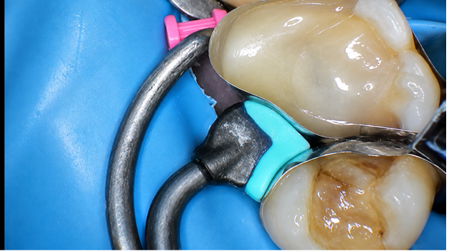
|
| Image 11: Tetric PowerFill Flow was first placed as a base, followed by Tetric PowerFill Composite, shade A |
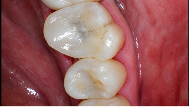
|
| Image 12: Restoration was then anatomically contoured and prepared for finishing and polishing |
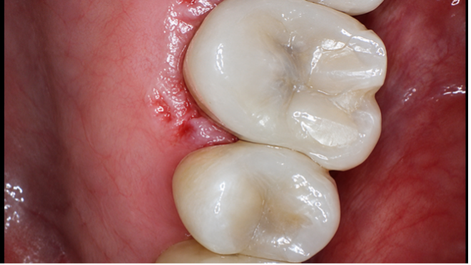
|
| Image 13: Postoperative view demonstrates a clearly defined palatal and gingival margin, providing a predictable foundation for the subsequent crown preparation |
Why DME Worked in This Case
The key advantage of DME was margin relocation.
Rather than immediately moving to surgical crown lengthening, the restorative margin was relocated coronally using bonded composite. This simplified access to the margin while preserving tooth structure and avoiding an additional surgical procedure.
The literature supports DME as a viable approach for selected localized subgingival defects, although clinicians should recognize that long-term clinical evidence is still limited.²,³
The technique is therefore less about the composite itself and more about control:
Control the field.
Control the matrix.
Control the adhesive interface.
Control the cervical contour.
When those four variables are controlled, the deep margin becomes much less intimidating.
Clinical Pearls
1. Isolate before you elevate.
DME is only as predictable as the isolation allows.
2. The matrix is everything.
A perfectly adapted DME band establishes the foundation for a clean cervical margin.
3. Respect the periodontium.
DME is not a substitute for surgical crown lengthening when the margin violates the periodontal attachment.
4. Don't underestimate the wedge.
Good wedging is essential to preventing gingival composite excess and establishing a clean cervical contour.
5. Total-etch is the gold standard.
A three-step etch-and-rinse system provides a well-established approach to dentin bonding and has substantial supporting laboratory and clinical literature.⁴–⁷
6. Think "margin relocation," not "box filling."
The objective of DME is to create a controllable restorative environment that makes everything that follows easier.
Conclusion
This case demonstrates a conservative restorative pathway for a fractured posterior cusp with a deep but manageable cervical margin.
By combining rubber dam isolation, soft-tissue management, DME, a three-step total-etch adhesive protocol, meticulous matrix adaptation and layered composite placement, the deep cervical margin was relocated to a more accessible position.
The immediate result was a clean, well-defined margin suitable for subsequent crown preparation.
DME doesn't eliminate the challenges of a deep margin—it moves the challenge to a place where we can control it.
Materials Used:
Reference
1. Miao C, Yang X, Wong MC, et al. Rubber dam isolation for restorative treatment in dental patients. Cochrane Database Syst Rev. 2021;5:CD009858. (PubMed)
2. Samartzi TK, Papalexopoulos D, Ntovas P, Rahiotis C, Blatz MB. Deep margin elevation: A literature review. Dent J. 2022;10(3):48. (PubMed Central (PMC))
3. Eggmann F, Ayub JM, Conejo J, Blatz MB. Deep margin elevation—Present status and future directions. J Esthet Restor Dent. 2023;35(1):26–47. (PubMed)
4. Van Meerbeek B, Yoshihara K, Yoshida Y, et al. State of the art of etch-and-rinse adhesives. Dent Mater. 2011;27(1):17–28. (PubMed)
5. De Munck J, Van Meerbeek B, Yoshida Y, et al. Four-year water degradation of total-etch adhesives bonded to dentin. J Dent Res. 2003;82:136–140.
6. De Munck J, Mine A, Poitevin A, et al. Meta-analytical review of parameters involved in dentin bonding. J Dent Res. 2012;91:351–357.
7. Peumans M, De Munck J, Mine A, et al. Twelve-year clinical evaluation of a three-step dentin adhesive. J Dent Res. Long-term clinical evidence supports the durability of three-step etch-and-rinse adhesive systems