FREE SHIPPING on orders over $475
FREE SHIPPING on orders over $475 | No Code Needed
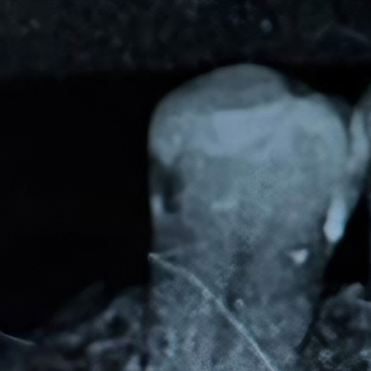
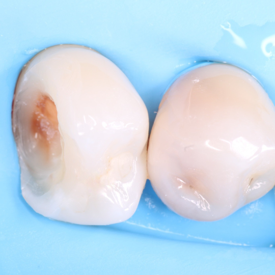
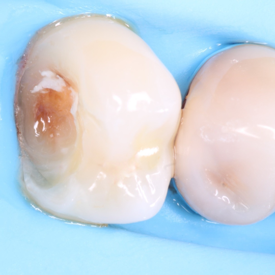
A 47-year-old female patient attended the dental practice concerned about the discoloration of a posterior tooth. The patient did not report any pain or sensitivity. Radiographic examination confirmed the presence of a mesial carious lesion on tooth #45.
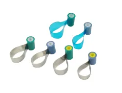
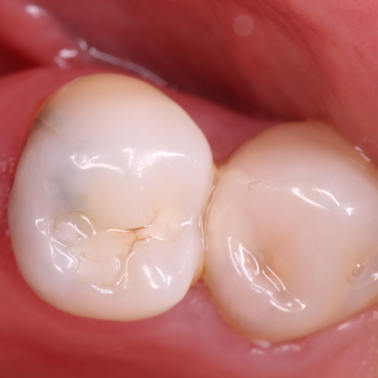
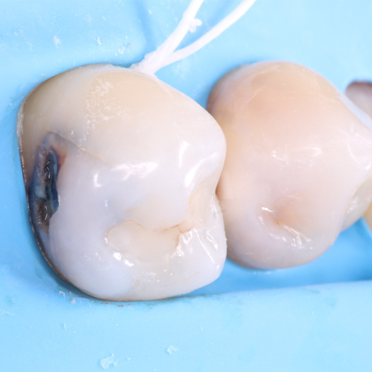
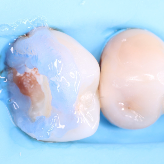
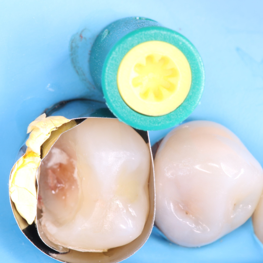
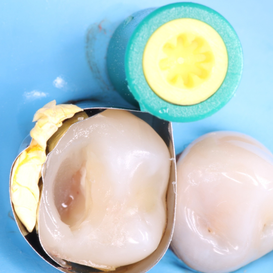
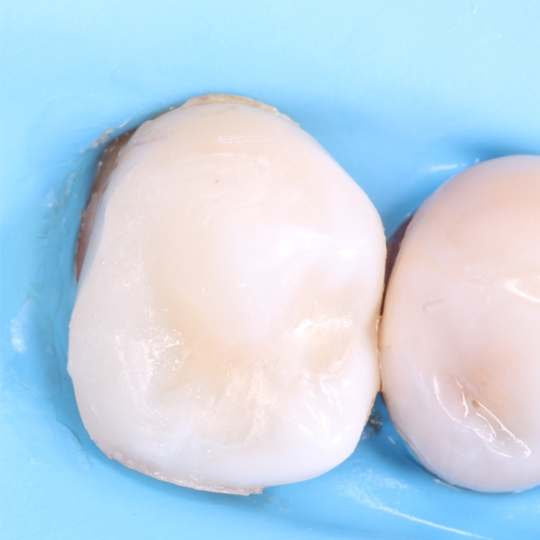
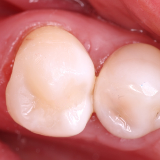
Clinically, a defective restoration and the presence of a carious lesion were observed on the occlusal surface. After local anesthesia, absolute isolation was performed using a Nic Tone medium rubber dam. The carious lesion was removed using a high-speed handpiece, followed by manual caries excavation with a dentin excavator. After cavity disinfection with 2% chlorhexidine, TheraCal was applied to the deepest area, closest to the pulp horn, and light-cured. Selective enamel etching was performed with 35% phosphoric acid for 15 seconds, followed by rinsing for twice the etching time and gentle drying. Two coats of universal adhesive were applied with active scrubbing, air-blown between each coat, and then light-cured. A 0.5 mm layer of flowable composite resin was placed on the floor and walls of the cavity and light-cured. The Garrison ReelMatrix™ matrix was then positioned, and a matrix-in-matrix technique was used to achieve better sealing. The proximal wall was built using the snowplow technique. Once the Class II cavity was converted into a Class I cavity, the restoration was completed using a horizontal incremental technique with Tetric N-Ceram A2, light-curing each increment with a curing unit delivering 1200 mW/cm². After rubber dam removal, high occlusal contacts were identified and adjusted. Finally, the restoration was finished and polished.
Tooth #44 will be restored at a subsequent appointment.
| 
| 
|
| 
| 
|
| 
| |
| 
| 
|
The use of the ReelMatrix™ system in clinical situations where an adjacent tooth is absent proves to be highly effective. Its adaptable matrix band provides an accurate fit around the tooth structure, allowing the clinician to properly reconstruct the proximal wall while closely reproducing the natural anatomical contour of the tooth. This facilitates improved restorative control, anatomical accuracy, and predictable clinical outcomes in challenging posterior restorations